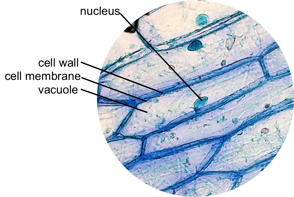
Onion epidermis, whole mount, 20x light micrograph. large epidermal Solved a) label each phase of mitosis for the onion root tip Onion bulb scale epidermis stock image. image of leaf
- Gold-plated onion cells make artificial muscles
Onion epidermal cells To prepare a stained, temporary mount of onion peel and to study its cells. Sample preparation process. (a) onion bulb. (b) bulb scale. (c) inner
Onion cells epidermal cell red epidermis peel wikipedia size
Epidermis de cebollaOnion cells light micrograph photomicrograph microscope high seen through cell resolution epidermis bulb organelles stock alamy wall scale Stomata leaf epidermis onion poresOnion 100x epidermis.
[diagram] onion epidermal cell labeled diagramOnion epidermal cell labeled diagram general wiring diagram Onion skin cell lpoOnion epidermis with large cells under microscope photograph by peter.
Epidermis epidermal muscles plated cell
Epidermis onion scale escala bulbo epiderme cebola cebolla bulb epidermide scala lampadina cipolla pilhasOnion epidermal cell diagram Biology practical| 9th biology practicalLm of cells in the epidermis of an onion.
Onion under skin stained epidermis mesophyll epithelium pee ling fig showing bulb mag microscopyPlant cell diagram Onion epidermal cell under microscope labeledHow many onion skins are there?.

Onion epidermis slide biology practical prepare 9th
Onion plant cell under microscope labeled onion cells onionCell onion wall structure plant function epidermis slide prepared flickr au yersinia veggies truth raw picture microscopes notes Onion peel temporary prepare stained epidermal microscope observation labelledOnion cells stock photos & onion cells stock images.
Onion epidermal cell labeled diagramOnion epidermal cell peel slide preparation practical experiment Onion epidermal nucleus zwiebel 100x hautzellen epidermis iodine magnification bulb alamy organelles epiderma showing nucleiOnion epidermal cells.

Onion leaf epidermis with stomata pores
Onion scale many labeled skins there microscopy evident every has magOnion epidermis 100x: general biology lab: loyola university chicago The inner epidermis of the onion bulb’s cataphylls (the onion skin).Onion cell cells microscope micrograph microscopic alamy stock magnification skin section high allium cepa mitosis root x100.
Onion epidermal cell- gold-plated onion cells make artificial muscles Onion epidermis[diagram] onion cell 40x labeled diagram.

Onion cells microscope hi-res stock photography and images
Cells cell plant vacuole onion microscope under structure microscopic do wall nucleus biology lab picture identify membrane epidermal diagram peelFun science blog: onion cell Onion epidermal cell labeled plasma membraneOnion cell epidermal diagram labeled cells microscope under drawing skin epidermis lab bulb mag membrane observation vacuole nucleus leaves requirements.
Onion epidermal drawings epidermis labeled biology chromosomes chromosome dna observation[diagram] onion epidermal cell labeled diagram What is the onion peel cell experiment? theory, video, procedureEpidermal cells mic epidermis.

Solved the next two images are real microscope views of the
Onion microscope epidermis furian hermes 26th .
.


Onion epidermis with large cells under microscope Photograph by Peter
- Gold-plated onion cells make artificial muscles

Onion Epidermal Cells | www.pixshark.com - Images Galleries With A Bite!

How many onion skins are there?

Plant Cell Diagram
![[DIAGRAM] Onion Cell 40x Labeled Diagram - MYDIAGRAM.ONLINE](https://i2.wp.com/medsci.indiana.edu/a215/oldvirtuallab/a215only/cell_cell_division/onion_root_tip/picture_labeled.jpg)
[DIAGRAM] Onion Cell 40x Labeled Diagram - MYDIAGRAM.ONLINE